


Despite efforts to develop treatments, there is still no cure for EVD. But scientists may have finally found a way to stop the virus from replicating.
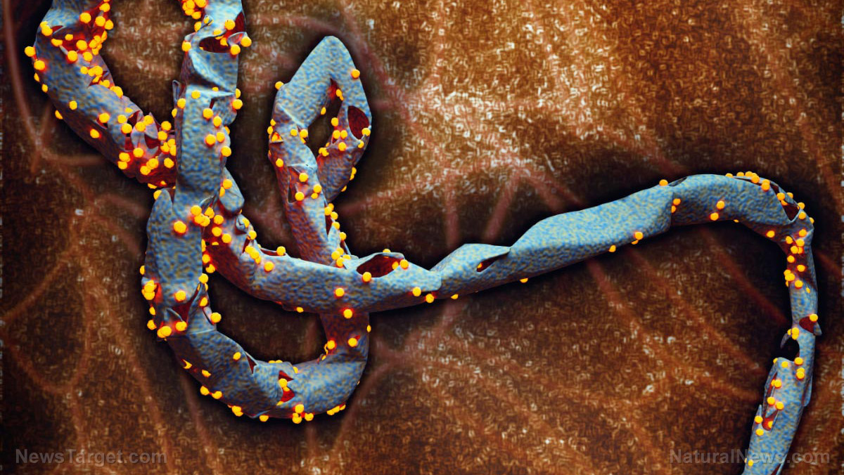
Robert Stahelin, a professor of medicinal chemistry and molecular pharmacology at Purdue University in Indiana, has been studying Ebola virus genes for years. In his latest research, Stahelin focused on a specific protein used by the Ebola virus for budding. Called Viral Protein 40 (VP40), this component of the virus' peripheral membrane is what allows it to cross the plasma membrane of human cells and replicate.
Together with researchers from the University of Notre Dame, Florida International University and The Scripps Research Institute in California, Stahelin looked at how VP40 interacts with molecules in the human cell membrane, particularly a membrane lipid known as phosphatidylserine (PS). They found that modifying a specific part of VP40 reduces its interaction with PS, which results in the Ebola virus being unable to form a viral envelope. The viral envelope plays an important role in infection as it allows the virus to enter its target cells, release its genetic content and package newly formed viral particles.
The role of VP40 during viral infection
Unlike living organisms, viruses are incapable of performing life-sustaining functions or reproduction. In order to replicate, they need to take over or "infect" units that are well-equipped to execute biological processes, such as cells. Viral infection of a cell involves several steps, beginning with the attachment to the cell membrane and ending with the release of newly formed viral particles. Cells that have receptors for specific viral proteins become susceptible to infection.

One distinct feature of Filoviruses like Ebola is that they have lipid envelopes. These viruses take those lipids from their host cells during infection. In the case of Ebola, Stahelin and his team found that VP40 makes up the layer beneath the virus' lipid envelope, suggesting that the protein is responsible for hijacking and taking the host lipid membrane to form the long viral envelope.
When the researchers looked at VP40's interaction with PS in the cell membrane, they discovered the specific part of the protein that binds with the lipid: a cationic patch at the end of an amino acid chain. They believe that this site is what controls the ability of VP40 to form a lipid envelope, which also serves as the virus' protection from the outside environment. (Related: Ebola transmission by aerosols confirmed: virus survives for days outside infected hosts.)
Changing the structure of VP40 weakens the Ebola virus' ability to replicate
VP40, being a transformer protein, is capable of rearranging itself into different structures. In their study, Stahelin's team found that monomeric, dimeric and octameric forms of VP40 bind to PS differently. Dimeric VP40 showed the greatest affinity to PS-containing membranes and was better at facilitating the replication of Ebola virus than monomeric VP40. On the other hand, VP40 binding to PS decreased by nearly tenfold in its octameric form.
“It’s exciting to learn that these different oligomeric structures bind differently with the human lipid cells. That might explain why there are different roles for this protein in the viral replication cycle,” said Stahelin.
Besides the molecular structure of VP40, the researchers also tried to modify its chemical structure. They replaced the hydrophilic (water-attracting) residues at the site where it binds to PS with alanine, a hydrophobic (water-repelling) amino acid. The hydrophilic residues are critical for viral entry into a cell and budding -- the process that enables a virus to exit its host. The loss of hydrophilic residues reduced VP40 binding to PS by 40-fold and stopped localization of the virus to the plasma membrane.
“This helps us understand how the virus uses human cell membranes to replicate and form new virus particles,” Stahelin said of their finding. “The virus needs this one lipid to form the new particle and infect other cells.”
This study was published in the Journal of Biological Chemistry.
Sources include:


Please contact us for more information.

